


CTA Head/Neck Images from Locked-In Case

A few months ago, I asked the plaintiff’s attorney for the Locked-In Syndrome case if he would be willing to send me the actual CTA images.
The patient and his family thought about it and agreed to send me the DICOM files so that I could share them with you all.
A chiropractor had adjusted the patient’s neck, immediately followed by severe dizziness and seizure-like activity.
The ER doctor ordered a CTA head/neck within minutes of arrival. The indication for study was “pain/dissection”.
Review the images at this link: https://www.dicomlibrary.com/meddream/?study=1.3.6.1.4.1.44316.6.102.1.20250620142526955.94039209153983692599
Note: Images load a bit slowly the first time through. They should scroll smoothly once you’ve looked through them the first time. DICOM Library was the best option I could find to upload the images and create a shareable link.
The radiologist wrote the following report:


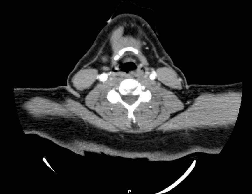
I’m not a radiologist, but here’s what stands out to me:
I agree with the radiologist that the right vertebral artery is much smaller caliber than the left. I lose track of it at the level of C1/C2 due to artifact around series 2, images 78-84, but don’t see it contributing to the basilar artery at all.
The left vertebral artery looks fairly normal to me throughout its course, although in the area of the same C1/C2 artifact there are a few images in which it appears much more narrow.
The most worrisome thing to me is that I lose the basilar artery by series 2, images 101 and it doesn’t reappear until image 108. A basilar obstruction is the finding that would make most sense in a patient with locked-in syndrome.
The expert witness radiologist felt that the left vertebral was dissected from C1/C2 all the way to the basilar artery, and also notes the high-grade basilar obstruction.

To be frank, I don’t think I would have seen the left vertebral dissection on my own.
The dwindling caliber of the basilar is easy enough for anyone to see, but the left vert dissection is very subtle to my eye.
The narrow caliber of the right vertebral artery is certainly easier to identify, and may have distracted from the findings on the left side.
I wonder if the patient’s vertebral artery anatomy explains why he had such a severe initial response to the dissection in the chiropractor’s office. His posterior circulation may have been dependent on the left-dominant vertebral artery, he had very little collateral flow through the right side at baseline, so reduction in left-side flow was especially catastrophic. In a patient with normal anatomy, the initial presentation may have been more subtle due to normal contralateral flow.
In my opinion, any criticism about the read on the vertebral arteries is secondary to the criticism of the missed basilar occlusion.
Become a better doctor by reviewing med mal cases.
Paying subscribers get a new case every week.
This case is an excellent opportunity for those of us who are generalists to review what posterior circulation pathology looks like on CTA.
Most generalists get comfortable with obvious findings like a saddle PE on a CTA chest, a kidney stone on a non-con CT abd/pelvis, or a head bleed on non-con head CT.
However, CTA head/neck are much more challenging studies to read, so many generalists don’t put as much effort into looking at them and rely more heavily on the radiology report.
I’d encourage you to spend some time reviewing the anatomy and looking through multiple examples of vertebral dissections and basilar occlusions.
If you can’t answer off the top of your head what artery the vertebrals originate from, what level they usually enter the vertebral column, describe their course at the C1/C2 level, or point to them on a CTA, it’s time to refresh your knowledge. You passed tests on this stuff in med school, but sometimes it fades in your memory and needs to be reviewed.
It’ll make you a better doctor and might even prevent a bad outcome like this.
The patient and his family have also released the head CT done in the ED, as well as the MRI done the following day.
CT head wo - https://www.dicomlibrary.com/meddream/?study=1.3.6.1.4.1.44316.6.102.1.202506202345569.5374284604742131407193
If you’re a radiologist, I’d love to hear your comments about the CTA.
Please leave a comment and teach us something!
Neuroradiologist here. There is quite a bit of streak artifact obscuring the distal vertebral arteries as well as some venous contamination and while the interpreting radiologist could be given some slack for having not detected the distal left vertebral artery dissection, what is not obscured is the basilar artery that is clearly occluded through much of its proximal extent- an unfortunate missed finding that should have been made. As a radiologist, I cannot say whether or not it would have made a difference in the patient's outcome so I would defer to the expertise of others to speculate on that.
Also, while I agree you could make a case for possible right vert dissection, the transverse foramina on the right are small indicating this is likely just a normal variant, congenitally diminutive artery.
Reviewed all the images and the cases you linked in Radiopedia- better ER doc for it now. Immense gratitude to the family for sharing the images of their loved for my learning. Disregarding my own opinions about how the case went (and chiropractors), the family has probably been through some awful times with all this.